กายวิภาคและเนื้อเยื่อวิทยาของต่อมย่อยอาหารในหอยนางรมปากจีบ Saccostrea cucullata (Born, 1778)
Main Article Content
Abstract
Methiya Uthamka, Supatta Chueycham, Kullanist Thanormjit and Sutin Kingtong
รับบทความ: 23 กุมภาพันธ์ 2560; ยอมรับตีพิมพ์: 15 พฤษภาคม 2560
บทคัดย่อ
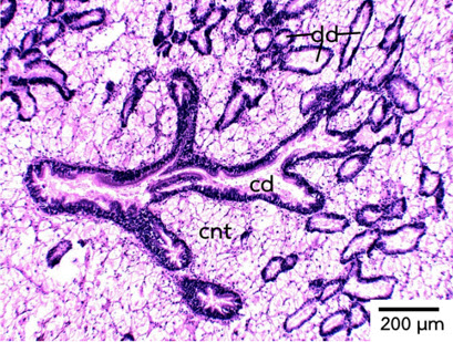
งานวิจัยครั้งนี้มีวัตถุประสงค์เพื่อศึกษากายวิภาคและเนื้อเยื่อวิทยาของต่อมย่อยอาหารในหอย นางรมปากจีบ Saccostrea cucullata (Born, 1778) ซึ่งเป็นสัตว์เศรษฐกิจที่มีการเพาะเลี้ยงในพื้นที่ชายฝั่งทะเลภาคตะวันออกของประเทศไทย โดยใช้วิธีการผ่าตัดเนื้อเยื่อและเทคนิคมิญวิทยา พบว่า ต่อมย่อยอาหารของหอยนางรมปากจีบ เป็นอวัยวะภายในที่อยู่ล้อมรอบกระเพาะอาหารและมีท่อเชื่อมต่อกับกระเพาะอาหาร เนื้อเยื่อของต่อมย่อยอาหารประกอบด้วยถุงปลายตันจำนวนมากแทรกอยู่ในเนื้อเยื่อเกี่ยวพัน แต่ละถุงมีขนาดเส้นผ่านศูนย์กลางประมาณ 70 – 100 µm ถุงปลายตันเหล่านี้เชื่อมต่อเข้ากับท่อรวมเพื่อเข้าสู่กระเพาะอาหาร ภายในถุงปลายตันประกอบด้วยเนื้อเยื่อบุผิวชั้นเดียว สามารถจำแนกเซลล์ที่พบได้ 2 ชนิด ตามรูปร่างของเซลล์และคุณสมบัติของการติดสีย้อม ได้แก่ เซลล์ย่อยอาหารเป็นเซลล์ทรงสูงและพบไซโทพลาซึมติดสีฮีมาท็อกไซลินจาง และเซลล์แบโซฟิลิกหรือเซลล์คริปท์ เป็นเซลล์ทรงเตี้ยและพบไซโทพลาซึมติดสีฮีมาท็อกไซลินเข้ม สำหรับท่อรวมของต่อมย่อยอาหาร พบเซลล์เยื่อบุผิวแบบหลายชั้นเทียมแบบมีซิเลียและพบเซลล์สร้างเมือกจำนวนมากแทรกอยู่ภายในเนื้อเยื่อบุผิวของท่อรวม ภายในเซลล์สร้างเมือกพบแกรนูลขนาดเล็กจำนวนมากย้อมติดสีอีโอซินเข้ม คาดว่าเป็นแกรนูลของเมือกที่จะหลั่งเข้าสู่ลูเมนเพื่อหล่อลื่นอาหารที่ผ่านเข้าและออกระหว่างกระเพาะอาหารและต่อมย่อยอาหาร
คำสำคัญ: หอยนางรมปากจีบ ต่อมย่อยอาหาร เซลล์ย่อยอาหาร เซลล์คริปท์
Abstract
The main objectives of current work was to investigate anatomical and histological structures of digestive diverticula in the Hooded-oyster, Saccostrea cucullata (Born, 1778), a commercially important species cultivated in the Eastern coast of Thailand, by dissection and histological techniques. The results showed that digestive diverticula of the Hooded-oyster is located in visceral mass and surrounding the stomach and connected to the stomach via collecting duct. The digestive diverticula compose of numerous blind-ending tubules, with the size of around 70 – 100 µm in diameter, distributed in connective tissue. These blind-ending tubules communicate with stomach by collecting ducts. The blind-ending tubules compose of single layer epithelium which consist of 2-cell types based on cellular characteristics and staining properties. These include digestive cell which is a columnar shape with pale hematoxylin staining in its cytoplasm and basophilic or crypt cell which exhibits a shot pyramidal shape with dark hematoxylin staining in its cytoplasm. The collecting ducts compose of pseudostratified columnar epithelium. A number of mucous or goblet cells are also distributed in the epithelia of the ducts. Within goblet cells, mucous granules are abundant which stained to the eosin. These granules are expected to secrete mucous into the lumen to lubricate epithelial surface and support food particle movement in the lumen.
Keywords: Oyster, Digestive diverticula, Digestive cell, Crypt cell
Downloads
Article Details

This work is licensed under a Creative Commons Attribution-NonCommercial 4.0 International License.
References
Arrighetti, F., Teso, V., and Penchaszadeh, P. E. (2015). Ultrastructure and histochemistry of the digestive gland of the giant predator snail Adelomelon beckii (Caeno gastropoda: Volutidae) from the SW Atlantic. Tissue Cell 47(2): 171–177.
Galtsoff, P. S. (1964). The American oyster Crassostrea virginica Gmelin. Fishery Bulletin. USA: Bureau of Commercial Fisheries.
Gros, O., Frenkiel, L., and Aldana, D. A. (2009). Structural analysis of the digestive gland of the queen conch Strombus gigas Linnaeus, 1758 and its intracellular parasites. Journal of Molluscan Studies 75(1): 59–68.
Mcelwain, A., and Bullard, S. A. (2014). Histo- logical atlas of freshwater mussels (Bivalvia, Unionidae): Villosa nebulosa (Ambleminae: Lampsilini), Fusconaia cerina (Ambleminae: Pleurobemini) and Strophitus connasaugaensis (Unioninae: Anodontini). Malacologia 57(1): 99–239.
Morton, B. (1996). The biology and functional morphology of Minnivola pyxidatus (Bivalvia: Pectinoidea). Journal of Zoology 240(4): 735–760.
Owen, G. (1973). The fine structure and histochemistry of the digestive diverticula of the protobranchiate bivalve Nucula sulcata. Proceeding of the Royal Society of London B 183: 249–264.
Spiers, Z. B. (2008). The identification and Distribution of an Intracellular Ciliate in Pearl Oysters, Pinctada maxima (Jameson 1901), Doctoral dissertation, Department of Veterinary and Biomedical Sciences, Murdoch University of Australia.
Taieb, N., and Vicente, N. (1999). Histochemistry and ultrastructure of the crypt cells in the digestive gland of the Aplysia punctada (Cuvier, 1803). Journal of Molluscan Studies 65(4): 385–398.
Yonge, C. M. (1926). XV.–The digestive di-verticula in the Lamellibranchs. Trans-actions of the Royal Society of Edinburgh 54(3): 703–718.
Khondee, P, Srisomsap, C., Chokchaicham-nankit, D., Svasti, J Simpson, R. J., Kingtong, S. (2016). Histopathological effect and stress response of mantle proteome following TBT exposure in the Hooded oyster Saccostrea cucullata, Environmental Pollution 218: 855–862.
